Decoding cancer cell by cell
A high-resolution map of the tumour
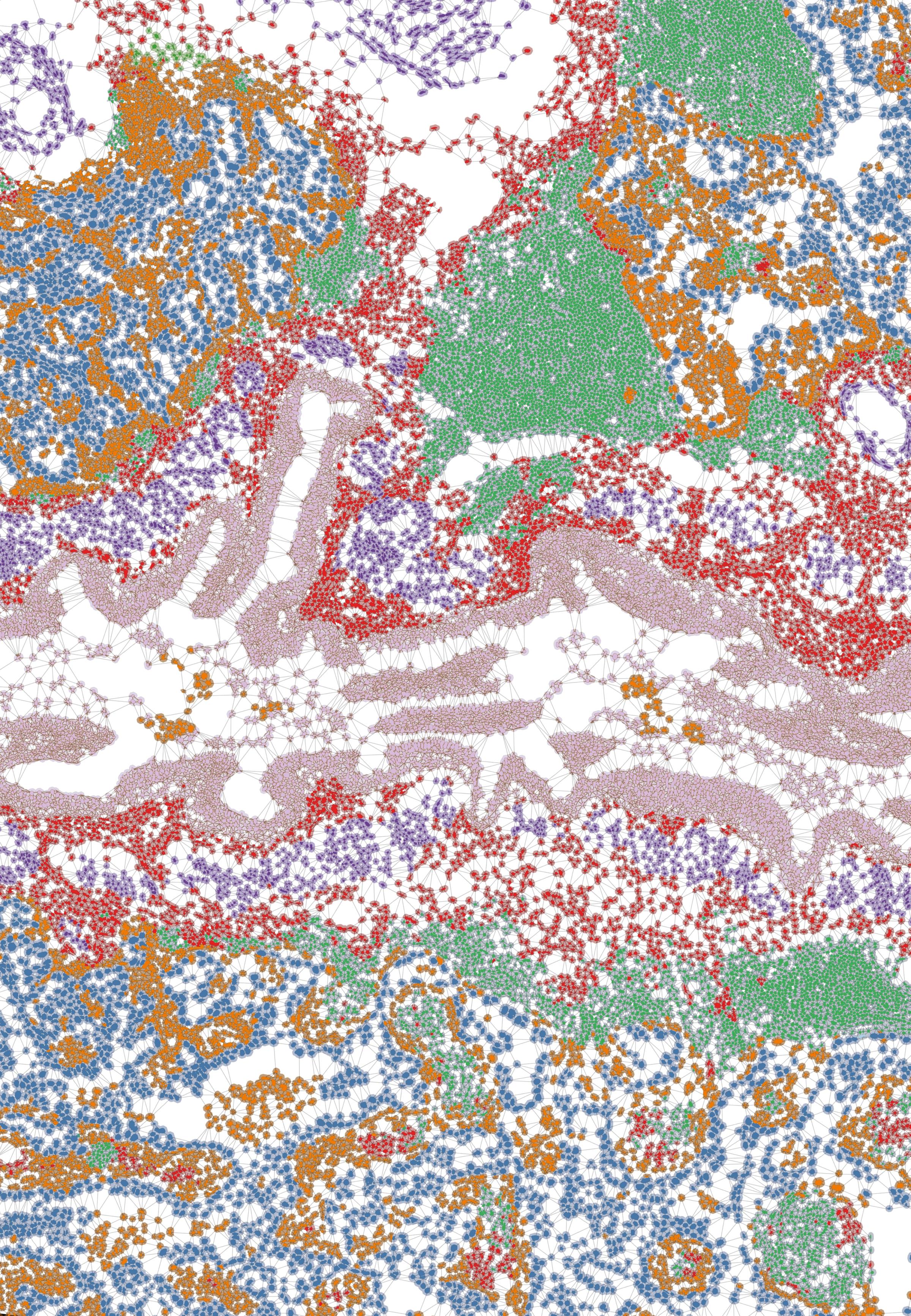
Generated by algorithms, cell-by-cell mapping of tumours reveals complex cellular profiles that are sometimes invisible using conventional methods. They are powerful tools for understanding tumour dynamics, anticipating resistance to certain treatments and identifying new therapeutic targets.
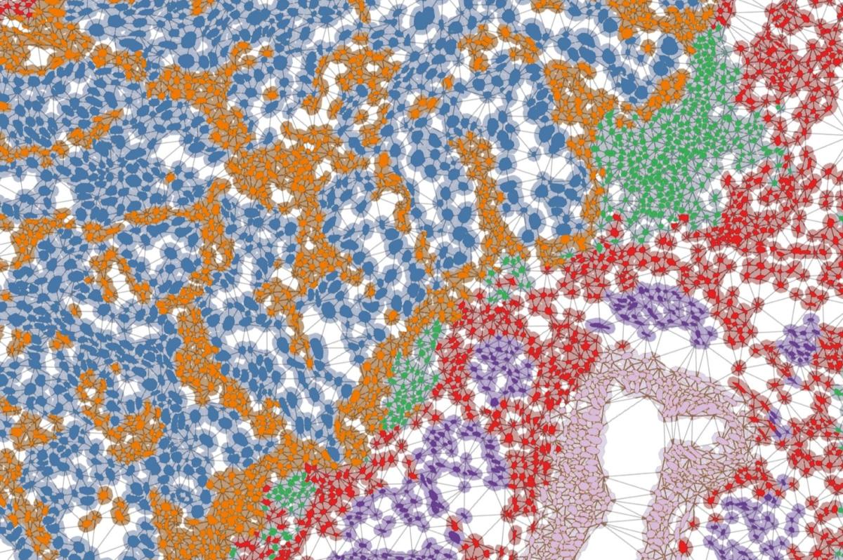
Tumours are actually mosaics of thousands of cells.
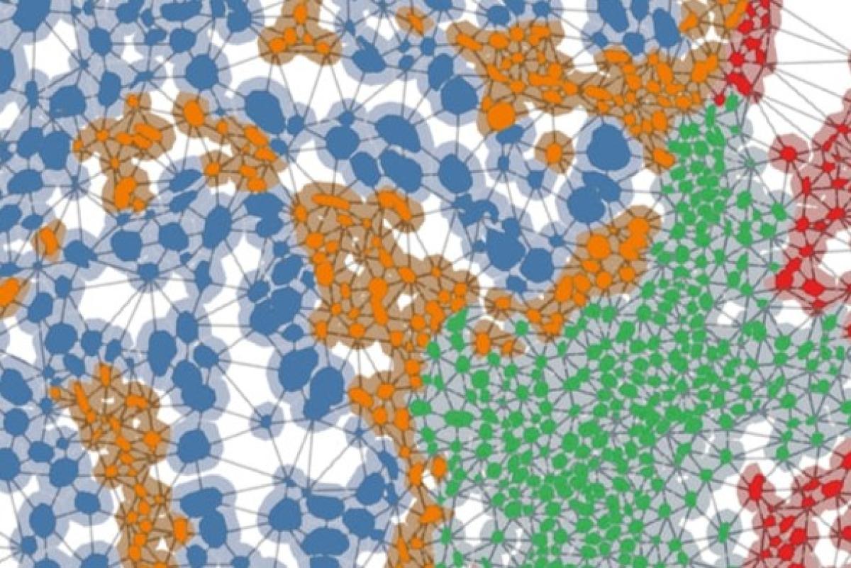
Each point is a cell, and each line an interaction.
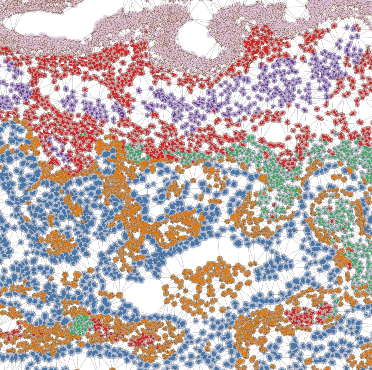
Each colour indicates a different type of cell:
Blue: cancer cells
Green: immune cells (T and B lymphocytes) that have infiltrated the area to fight the cancer cells
Red: fibroblasts and stromal cells forming the elastic framework of the lungs
Purple: peribronchial muscle cells regulating the diameter of the bronchi
Pink: ciliated epithelial cells and basal cells responsible for filtration and renewal of lung tissue
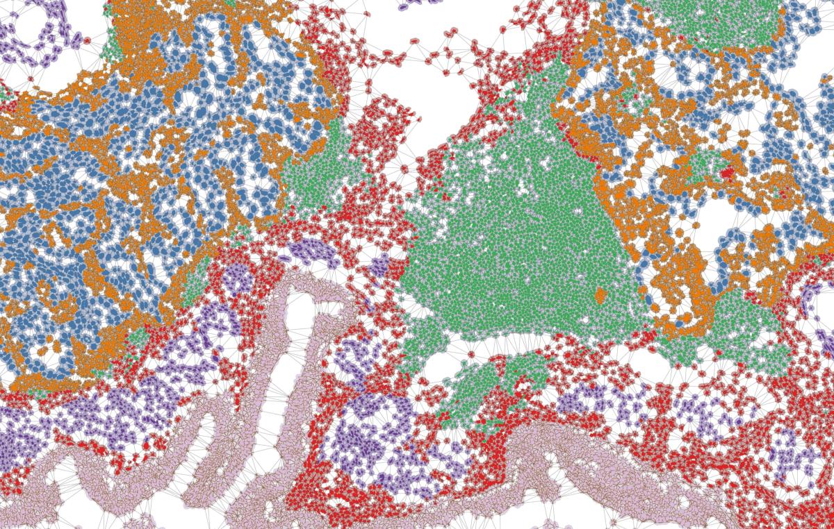
On this lung cancer section, more than 10,000 cells were analysed and classified using bioinformatics.